
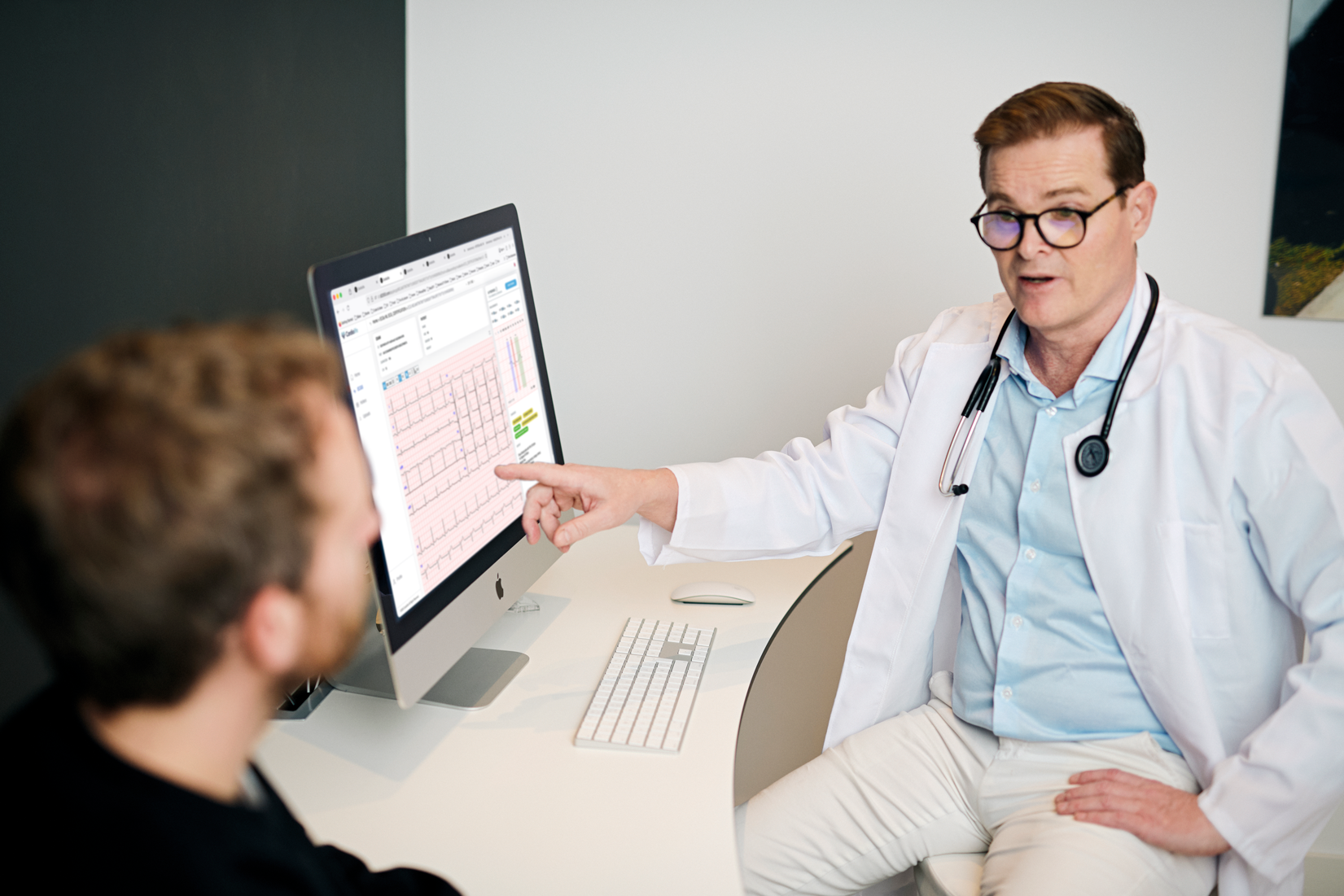
From ECG signal
to clinical insight
in seconds
AI that automates the full ECG workflow for fast, accurate, and consistent diagnostics.
Proven Clinical
Accuracy
Expert-level interpretation trained on 1,26 million ECGs, achieving up to 100% balanced accuracy in clinical validation.

Device-Agnostic
Compatible with a broad range of widely used ECG formats and supported devices for practical interoperability.

Multilingual Support
Available in English, Portuguese, French, German, Italian and Spanish.

Broad Diagnostic Coverage
Expert-level rhythm and morphology detection across 34 supported ECG findings delivered in seconds. These include all the main conduction, structural and rhythm disorders.

Seamless Integration
Integrates smoothly via cloud or API into existing systems, enabling fast adoption with minimal workflow disruption.

Certified & Compliant
Fully MDR-certified and aligned with international standards for trusted clinical use.
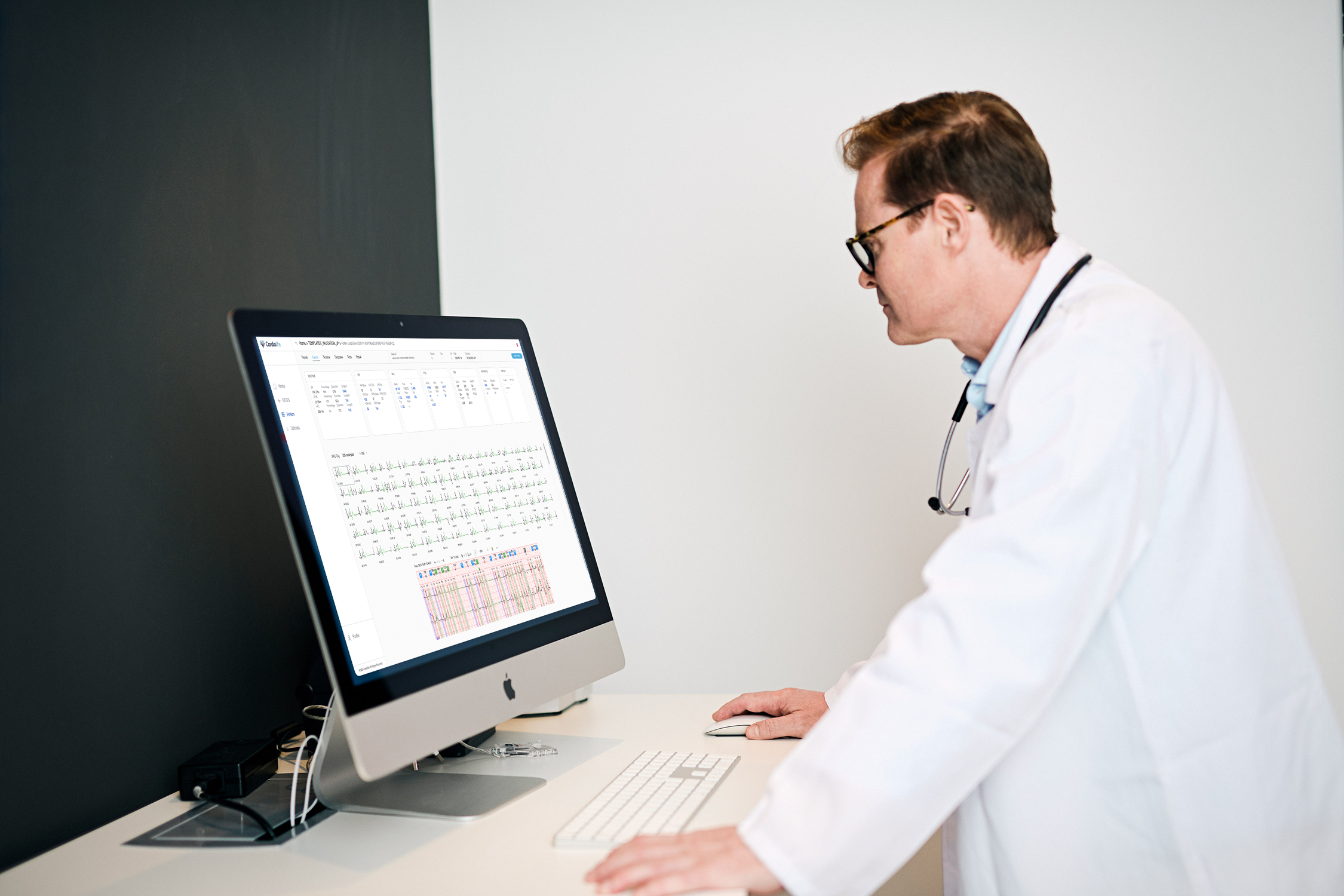
Long-Duration ECG Support
Analyze ECGs of any duration and lead configuration — from standard 12-lead resting ECGs to long-term Holter recordings, single- or multi-lead.

Editable Reports
Transparent, clinician-editable outputs for personalized, seamless review and adjustment.
940,000 patients
50 + healthcenters
1.260,000 ECGs
Fast, accurate ECG interpretation with
an all-in-one clinical workspace
View, review, edit and report ECGs with AI-driven insights built for real-world clinical practice.
1
Expert-level rhythm and morphology detection across 34 supported ECG findings in seconds.
ECG Findings Overview
2
Automatic beat segmentation and precise measurement of intervals, axis and morphology measurements.
Metric Overview
4
A full-featured workspace with tools for waveform inspection, lead organisation and signal exploration.
ECG Workspace
3
Structured and editable, clinician-ready reports generated automatically from AI findings and metrics with EHR integration capability.
Fully Standardized Clinical Text Reports
5
Perform precise manual caliper measurements to verify findings and support clinical judgement.
Manual Measurements
High-Precision Detection
Covering 34 ECG findings, 17 Holter findings, and 7 advanced metrics — with up to 100% accuracy
-
SR Sinus Rhythm
Accuracy 92%SA Sinus Arrest/Sinus Pause
Accuracy: 94%PAC Premature Atrial Complex
Accuracy: 98%PACAB Premature Atrial Complex with Aberrant Conduction
Accuracy: 95%EAR Ectopic Atrial Rhythm
Accuracy: 95%AFIB Atrial Fibrillation
Accuracy: 100%AFL Atrial Flutter
Accuracy: 98%SVT Supraventricular Tachycardia
Accuracy: 98% (internal dataset performance test)PVC Premature Ventricular Complex
Accuracy: 100%VT Ventricular Tachycardia
Accuracy: 100%APACE Atrial Pacing
Accuracy: 98%VPACE Ventricular Pacing
Accuracy: 99%AVB1 1st Degree Atrioventricular Block
Accuracy: 97%AVB2M1 2nd Degree Atrioventricular Block Mobitz I
Accuracy: 97%AVB2M2 2nd Degree Atrioventricular Block Mobitz II
Accuracy: 90%AVB2:1 2nd Degree Atrioventricular Block 2:1
Accuracy: 97%AVB3 3rd Degree/Complete Atrioventricular Block
Accuracy: 90%WPW Wolff-Parkinson-White/Ventricular Pre-excitation
Accuracy: 99%IRBBB RSR’ pattern/Incomplete Right Bundle Branch Block
Accuracy: 95%RBBB Complete Right Bundle Branch Block
Accuracy: 100%LAFB Left Anterior Fascicular Block
Accuracy: 98%LBBB Left Bundle Branch Block
Accuracy: 100%LAE Left Atrial Enlargement
Accuracy: 94%LVH Left Ventricular Hypertrophy
Accuracy: 91%LVLL Low Voltages in the Limb Leads
Accuracy: 100%LVCL Low Voltages in the Chest Leads
Accuracy: 97%BRS Brugada Pattern Sindrome
Accuracy: 99%OMII Old inferior Myocardial Infarction
Accuracy: 92%OMIA Anteroseptal or anterior myocardial infarction old
Accuracy: 96%PRWP Poor R Wave Progression V1-V3
Accuracy: 97%ERP Early Repolarization Pattern
Accuracy: 91%STD ST Segment Deviation
Accuracy: 91%JTP Juvenile T-wave Pattern
Accuracy: 96%LVHST Left Ventricular Hypertrophy with ST-T Changes
Accuracy: 96%
Accuracy is presented as balanced accuracy, defined as the average of sensitivity and specificity.
All diagnoses meet or exceed the acceptance criteria of 90%
-
SR Sinus Rhythm
Accuracy: 94%SP Sinus Pause
Accuarcy: 94%PAC Premature Atrial Complex
Accuracy: 98%PACAB Premature Atrial Complex with Aberrant Conduction
Accuracy: 98%EAR Ectopic Atrial Rhythm
Accuracy: 95%AFIB Atrial Fibrillation
Accuracy: 100%AFL Atrial Flutter
Accuracy: 98%SVT Supraventricular Tachycardia
Accuracy: 98% (internal dataset performance test)VT Ventricular Tachycardia
Accuracy: 100%APACE Atrial Pacing
Accuracy: 98%VPACE Ventricular Pacing
Accuracy: 99%PVC Premature Ventricular Complex
Accuray: 100%AVB1 1st Degree Atrioventricular Block
Accuracy: 97%AVB2M1 2nd Degree Atrioventricular Block Mobitz I
Accuracy: 97%AVB2M2 2nd Degree Atrioventricular Block Mobitz II
Accuracy: 90%AVB2:1 2nd Degree Atrioventricular Block 2:1
Accuracy: 97%AVB3 3rd Degree Complete Atrioventricular Block
Accuracy: 90%
Accuracy is presented as balanced accuracy, defined as the average of sensitivity and specificity.
All diagnoses meet or exceed the acceptance criteria of 90%
-
R-R Time difference between two consecutive R points
ICC: 0,99HR – Heart rate (beats per minute), derived from RR interval (HR = 60/RR)
ICC: Not applicable (derived from RR)HRV – Heart rate variability, calculated from successive RR intervals
ICC: Not applicable (derived from RR)PQ (PR) Measures the time from the beginning of the P wave to the beginning of the QRS complex
ICC: 0,90QRS Representing ventricular depolarization
ICC:0,86QT Time from the beginning of the QRS complex to the end of the T wave
ICC: 0,85QRS Axis QRS Axis orientation (one of 4 values: normal, left, right, indeterminate)
ICC: 0,96
All quantitative metrics meet or exceed the acceptance criterion of ICC ≥ 0.85
Ready to enhance your ECG workflow with AI?
Discover how Cardiolife integrates seamlessly into your clinical ecosystem.